Sunday, March 9, 2008

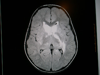
Acute Disseminated Encephalomyelitis (ADEM)


Acute disseminated encephalomyelitis (ADEM) is an immune mediated disease of brain. It usually occurs following a viral infection but may appear following vaccination, bacterial or parasitic infection, or even appear spontaneously. It involves autoimmune demyelination.Full recovery is seen in 50 to 75% of cases, ranging to 70 to 90% recovery with some minor residual disability, with an average time to recover of one to six months.MRI is highly sensitive in detecting white matter lesions and the lesions described are rather extensive and subcortical in location. Involvement of the deep gray matter, particularly basal ganglia, is more frequent. Use of high-dose methylprednisolone, plasma exchange, and IVIG are based on the analogy of the pathogenesis of ADEM with that of multiple sclerosis (MS). Differentiation of ADEM from the first attack of MS is important from prognostic as well as therapeutic point of view. This differentiation is more relevant to India where the incidence of MS is low.

To read more: click here

Dermoids Here and There


Intracranial Dermoids:
Most dermoid tumors have signal intensity characteristics similar to fat—that is, they are hyperintense on T1-weighted images and hypointense on T2-weighted images. Fat-suppression techniques may be helpful in confirming the presence of fat in the lesion. Centrally, dermoid tumors may appear inhomogeneous due to the presence of hair follicles, calcifications, and cellular debris.Rupture of a dermoid tumor can result in fat droplets in the subarachnoid spaces or ventricles.


Spinal Dermoids:
The most frequent site of these tumors is in an extramedullary location; a few may lie in the substance of the cord itself. Complications of the cyst include secondary infection, particularly when associated witha dermal sinus. Rarely these lead to chemical meningitis from the cholesterol-laden keratin released in the subarachnoid space from a ruptured cyst leading to a foreign body type of reaction.


Presacral Dermoid:
A unilocular cystic mass located at or near the midline, with signal intensity similar to that of fat, is highly suggestive of a dermoid tumor.

Submitted by Dr MGK Murthy (Elbit Medical Diagnostics)

Chordoma


Large expansile infiltrative lesion in the S1 and S2 vertebral bodies with pre and paravertebral and epidural components with consequent neural compromise and spinal canal narrowing.
Chordomas are rare tumors that arise from embryonic notochordal remnants along the length of the neuraxis at developmentally active sites.Chordomas are rare neoplasms. As primary intracranial neoplasms, they only constitute 0.2% of all CNS tumors. Chordomas generally occur in 3 locations, which are, in descending order of frequency, the sacrum, intracranially at the clivus, and along the spinal axis.CT scan or MRI studies are indicated to evaluate the extent of the tumor and to identify the tissues that the chordoma has infiltrated.The 5-year survival rate is estimated to be 51%, and the 10-year survival is estimated to be 35%. Factors that may improve prognosis are young age, complete resection, and the addition of radiation therapy in incompletely resected tumors.

You can find more information on this topic here

Saturday, March 8, 2008

Coexistent Carcinomas


Known case of prostatic carcinoma with increased PSA levels and osteoblastic metastasis in the spine. Interestingly this patient also had para-aortic adenopathy and a testicular swelling. This could well represent metastatic lymphadenopathy but the question of coexistent prostatic and testicular carcinomas should be raised. The incidence of coexistent malignancies is high in the elderly.

Malignant chacracteristics of a breast lesion


MRI - Sensitivity - 93% Specificity - 70%
1. Opacity
2. Enhancement pattern : patchy
- invasive: pheripheral or rim
- DCIS : linear
3. Type 3 time intensity curve
- early steep rise within 5 mts with a 70% increase in SI
- rapid washout
4. Tumour recurrence – radiation fibrosis does not enhance after 18 mts

Ultrasound :
Poorly reflective
Ill defined
Heterogenous internal echo pattern
Absent farwall echoes
Posterior acoustic shadowing
Taller than wide


Mammography:UOQ - 50% Retroareolar 18%
Opacity - stellate,spiculated,comet tail
- high density
- nonhomogenous
- wide halo
Asymmetric Density
Microcalcification – 30% of cases
- < 0.5 mm & > 5 particles per cc
- segmental, cluster distribution
- pleomorphic : linear, branching, punctate
- eccentric location in opacity