Friday, January 25, 2008

Glomus Jugulare


Large expansile grossly destructive irregular space occupying lesion involving the left temporal bone with intracranial extension and mass effect on brain stem, fourth ventricle and temporal horn.
Glomus jugulare tumors are rare, slow-growing, hypervascular tumors that arise within the jugular foramen of the temporal bone. They are included in a group of tumors referred to as paragangliomas, which occur at various sites and include carotid body, glomus vagale, and glomus tympanicum tumors.
The female-to-male ratio is 3-6:1. Glomus jugulare tumors have been noted to be more common on the left side, especially in females. The Glasscock-Jackson and Fisch classifications of glomus tumors are widely used. The Fisch classification of glomus tumors is based on extension of the tumor to surrounding anatomic structures and is closely related to mortality and morbidity.
Surgery is the treatment of choice for glomus jugulare tumors. Surgical approach depends on the localization and extension of the tumor. Intraoperative monitoring including EEGs and somatosensory-evoked potentials (SSEPs) are routinely used.Gross total resection of some extensive tumors may be extremely difficult and may carry unwarranted risk. In such cases, radiotherapy may be indicated to treat residual tumor following subtotal resection.

Submitted by Dr VN Goud (Elbit Medicla Diagnostics)

You can find more information on this topic here

T2 Relaxometry Secrets in Epilepsy Protocol

Hippocampal structural damage is equivocally depicted by spectroscopy. For diagnostic and pre-operative evaluation, hippocampal volumetry and T2-relaxometry provide maximal accuracy. Amygdala quantifications are irrelevant in the pre-operative evaluation but may be useful for diagnostic purposes. Of the three qMRI modalities tested, T2-relaxometry provided the best balance between diagnosis accuracy and time-efficiency to lateralize a sclerotic lesion on the majority of the patients. The salient points to rememberare:
- independent of magnetic strength, age, sex or side
- can be done not only in hippocampus but also in anterior temporal lobe, parietooccipiatal lobe, amgdela etc.
- complete symmetry exists between both sides (< 7 ms)
- normally should not exceed 90 to 97 ms
- if volume calculations and T2 values do not match believe the T2 values
- epileptogenic focus is the key to treatment

Submitted by Dr MGK Murthy (Elbit Medical Diagnostics)

Reference: click here

TB Meningitis


Multiple conglomerate ring enhancing lesions in the basal cisterns extending along tentorium predominantly on left side with lipid lactate peaks on MRS and dilated ventricular system.
The incidence of CNS TB is related to the prevalence of TB in the community, and it is still the most common type of chronic CNS infection in developing countries.
In an immunocompetent individual, CNS TB usually takes the form of meningitis that causes an acute-to-subacute illness characterized by fever, headache, drowsiness, meningism, and confusion over a period of approximately 2-3 weeks.
Two rare forms of TBM are serous TB meningitis and TB encephalopathy. Serous TB meningitis is characterized by signs and symptoms of a mild meningitis with spontaneous recovery. TB encephalopathy usually occurs in a young child with progressive primary TB; the presentation is that of reduced levels of consciousness with few focal signs and minimal meningism.

You can find more information on this topic here

Arteriovenous Malformation


Large AVM in right mid and superior perietal region with arterial feeders from right middle cerebral and part of anterior cerebral arteries and draining into superior sagital sinus and adjacent cortical veins.
An arteriovenous malformation (AVM) is a tangled cluster of vessels, typically located supratentorially, in which arteries connect directly to veins with no intervening capillary bed. A 1988 study of more than 500 patients showed that the core, or nidus, of a compact AVM was from 2-6 cm in diameter in 77%.AVMs account for approximately 11% of cerebrovascular malformations. AVMs are more likely than other types of vascular malformations to be clinically symptomatic. AVMs are categorized by their blood supply. Pial or parenchymal AVMs are supplied by the internal carotid or vertebral circulation; dural AVMs, by the external carotid circulation; and mixed AVMs, by both. A pediatric variant of AVM is the vein-of-Galen aneurysm, in which an AVM drains to and dilates the great vein of Galen.
The Spetzler and Martin grading system attempts to predict risk of surgical morbidity and mortality by assigning points to an AVM on the basis of its size, the eloquence of the adjacent brain, and the pattern of venous drainage.

You can find more information on this topic here

Thursday, January 17, 2008

Digital Breast Tomosynthesis


Tubulolobular adenocarcinoma seen on conventional digital mammogram (left) and in a slice from a tomosynthesis dataset (right).

Digital breast tomosynthesis boosts radiologists' cancer detection rates compared to digital mammography alone. The researchers conducted a multicenter, multi-reader study to evaluate reader performance with tomosynthesis.
It's done like a regular mammogram and still involves compressing the breast. But it takes a 3-D view with a CT scan in an arc around the breast. The machines are expensive, and as with many new techniques, testing will be expensive too. It only takes a few seconds to do the tomosynthesis part of the exam, but then the machine will also take a regular mammogram.

You can find more information on this topic here

Neurological Manifestations in Sjogren's Syndrome


Known case of Sjogren's Syndrome with hypothyroidism and hypertension.
Fairly large, symmetrical hyperintensities in periventricular frontal, parietal and parietooccipital white matter regions with similar smaller lesions in brainstem, cerebellum, bilateral thalamic and capsuloganglionic regions.
Possible cause is autoimmune pathology with vascular involvement. Corticosteroid therapy results in rapid and nearly complete resolution of the cortical lesions with marked improvement of the clinical manifestations. Memory disturbance is a rare initial manifestation in meningoencephalitis associated with SjS.

Ref: Journal of the Neurological Sciences, Volume 232, Issue 1-2, Pages 111-113

Submitted by Dr MGK Murthy (Elbit Medical Diagnostics)

Carcinoma Tongue


The most common type of cancer of the tongue is Squamous Cell Carcinoma. The oral tongue and the base of the tongue comprise the whole tongue but it is important to know that they develop from different embryonic tissue and really are somewhat dissimilar. Most importantly, this explains why the treatment for squamous cell carcinoma for the oral tongue is usually quite different from the treatment for squamous cell carcinoma of the base of tongue.
Unlike oral tongue cancers, base of tongue squamous cell cancer is usually larger when diagnosed because in the early stages it can not be seen and it creates few, if any, symptoms. Later however, base of tongue cancer may create pain, a sense of fullness, changes in what the voice sounds like, and perhaps even some difficulty in swallowing. Also, because the diagnosis often comes a bit later, a greater number of patients with this disease will already have neck metastasis in the lymph nodes of the neck, by the time they are seen by the Head and Neck Surgeon.

You can find more information on this topic here

Submitted by Dr Kiran David (Elbit Medical Diagnostics)

Wednesday, January 16, 2008

Cistern of Great Cerebral Vein


This patient presented with recurrent seizures.
The cistern of great cerebral vein occupies the interval between the splenium of the corpus callosum and the superior surface of the cerebellum; it extends between the layers of the tela chorioidea of the third ventricle and contains the great cerebral vein.Synonyms of cistern of great cerebral vein include: ambient cistern, Bichat's canal, Bichat's foramen, superior cistern.

TB of Lower Cervical Spine


This patient complained of pain in the neck. Plain radiography of the cervical spine did not show anything. Chest x-ray showed features of pulmonary TB. MRI showed the extensive involvement of the spine.
The pattern of cervical tuberculosis in children differs significantly from that in adults. This is related to the relative immunity of adults to the disease.Pain is a dominant feature in tuberculosis of the cervical spine.In the “adult” type the disease is much more localised and produces less pus. Often in Pott’s paraplegia the cord is compressed from its anterior aspect.Operative relief, if attempted, should therefore also be from the front.

Case submitted by Dr Kiran David (Elbit Medical Diagnostics)

You can find more information on this topic here

Saturday, January 5, 2008

Perfusion Explained


Dynamic susceptibility contrast perfusion imaging:
-rapid injection of MR contrast agent
-intravascular T2 relaxation with signal loss
-dynamic time-signal cure on each pixel
Three major perfusion parameters:
-negative enhancement integral = cerebral blood flow (CBV)
-mean time to enhance (MTT)
-blood flow CBV/MTT
Perfusion imaging:
-differentiate ischaemic stroke from haemorrhagic stroke
-more sensitive than diffusion imaging
-candidates for anti-coagulant therapy detected earlier
-determines the salvagable brain tissue
-reduces the susceptibility of brain to further ischaemic damage

You can find more information on this topic here

Coccygodynia


Common causes: anorectal infection, chronic trauma involving poor sitting posture, acute trauma, and parturition.
The pain originates in spasm of the levator ani and coccygeus (and at times in the mesial fibers of the gluteus maximus) muscles.
X-ray examination is of diagnostic value only in cases of suspected fracture or dislocation. MRI is the investigation of choice.
Relief obtained by injections of alcohol, or of various anesthetic agents, with or without cortisone.
Treatment by properly applied massage of the levator ani and coccygeus muscles, and at times of the mesial fibers of the gluteus maximus muscle gives relief.

You can find more information on this topic here

Friday, January 4, 2008

Pontine Glioma


Brainstem gliomas occur at 3 distinct anatomic locations—diffuse intrinsic pontine, tectal, and cervicomedullary. Intrinsic pontine gliomas carry a grave prognosis. Brainstem gliomas have been reported to make up 2.4% of all intracranial tumors in adults and 9.4% of intracranial tumors in children.Morbidity is due to the location of the space-occupying lesion and compression of surrounding structures; because these structures regulate basic body functions of blood pressure, respiration, and swallowing as well as motor and sensory functions, compression can produce substantial neurological disability. Bimodal age distribution has been noted, with a peak incidence in the latter half of the first decade of life and a second peak in the fourth decadeThe typical MRI appearance of a brainstem glioma is an expansile, infiltrative process with low-to-normal signal intensity on T1-weighted images and heterogeneous high-signal intensity on T2-weighted images, with or without contrast enhancement.

To read more: click here

Fluid Levels in Medical Imaging

Radiologists must be familiar with the appearances and significance of fluid levels. It is important to appreciate that only images in the vertical plane will show this phenomenon.On radiographs the common fluid levels : chest (achalasia, hiatus hernia, hydropneumothorax or cavitating lesion), abdomen (intestinal obstuction) and truama (lipohaemarthrosis in knee and paranasal sinuses in facial fractures).CT with its high inherent contrast between gas, fat and fluid is an excellent technique for the demonstration of fluid levels in abdomen, chest and in extremity truama. MRI adds greater inherent contrast for different types of liquids and is used to demonstrate layering in lipohaemarthrosis, staging bone and soft tissue tumours, aneurysmal bone cyst, giant cell tumour and synovial sarcoma. Although ultrasound is the most difficult imaging technique to reliably demonstrate fluid levels it can be used to show levels due to seperation of blood withing a false aneurysm.

To read more: Clinical Radiology December 2007

Lead Poisoning


The salient manifestations of lead poisoning:
1.Dense metaphyseal lines
2.Lead encephhalopathy:
Acute – cerebral oedema & microhaemorrhages
Chronic – patchy calcifications, atrophy & white matter changes
3.Sutural widening
4.Hepatospleenomegaly
5.Abdominal radiographs may show lead containing objects in GIT

Thursday, January 3, 2008

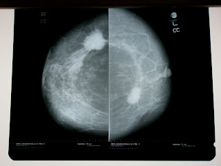
Multicentric Carcinoma Breast


Clinical Issues:
Multicentric in 10% of cases
Second focus may be microinvasive
Axillary positivity is 52%
Directly proportionate to tumour volume
Evaluation of each lesion necessary

Cerebral Venous Thrombosis


Cerebral venous thrombosis is a rare condition affecting predominantly adolescents or young adults. The presentation is often non-specific, and delay in diagnosis is common.This case draws attention to the multi-causal nature of cerebral venous thrombosis in young adults, and highlights the issue of masked mastoiditis. A coordinated approach by otolaryngological and haematological teams is recommended in such cases.