Tuesday, February 19, 2008

Local Gigantism of Right Ankle and Foot


Large diffuse irregular hypertrophy of the soft tissues involving the distal leg, ankle and foot on the right side. No evidence of phleboliths or signs of arteriovenous fistulae or fat elements. The differential here for local gigantism would include Klippel Trenaunay Syndrome and Plexiform Neurofibromatosis.
Klippel-Trenaunay syndrome (KTS) is defined by the presence of a combined vascular malformation of the capillaries, veins, and lymphatics, congenital venous abnormalities, and limb hypertrophy. Most patients with KTS can be treated conservatively with compression stockings or pneumatic pumps. Compression stockings decrease edema, act as a barrier for minor trauma, and reduce venous insufficiency.
Surgical Care - Servelle reported successful surgical intervention (resection or ligation of abnormal blood vessels) in more than 700 patients with KTS. Most medical centers have tried to avoid surgical intervention. Surgical treatment can be complicated by infection, lymph seepage, and skin breakdown. Intravenous sclerotherapy has been proposed as an alternative to surgical intervention in KTS and to embolization in PWS. Reports exist of the use of a sclerosant in microfoam.
Clinicians at all centers agree that a leg length discrepancy of more than 2.0 cm warrants epiphysiodesis. Hypertrophied digits with severe deformity and infection may require amputation.

You can find more information on this topic here

Posterior Reversible Encephalopathy Syndrome (PRES)


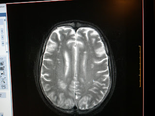
Multiple illdefined irregular altered signal intensity areas involving both parietoccipital regions in parasagital location.
Posterior Reversible Encephalopathy Syndrome (PRES) or Reversible Posterior leukoencephalopathy syndrome (RPLS) is an increasingly recognized neurologic disorder with characteristic computed tomographic (CT) and magnetic resonance (MR) imaging findings, and it is associated with a multitude of diverse clinical entities. These include acute glomerulonephritis, preeclampsia and eclampsia, systemic lupus erythematosus, and thrombotic thrombocytopenic purpura and hemolytic-uremic syndrome, as well as drug toxicity from agents such as cyclosporine, tacrolimus, cisplatin, and erythropoietin. Most, but not all, cases manifest with acute to subacute hypertension, and seizures are also frequent. Classic CT findings are those of bilaterally symmetric low attenuation in the posterior parietal and occipital lobes, whereas MR imaging demonstrates hyperintensity on T2-weighted images in the same distribution.
Two pathophysiologic mechanisms for RPLS have been proposed. One postulates cerebral vasospasm with resulting ischemia within the involved territories, whereas the other posits a breakdown in cerebrovascular autoregulation with ensuing interstitial extravasation of fluid. Diffusion MR imaging can be used to discriminate between these two possibilities, as the cytotoxic edema of cerebral ischemia demonstrates decreased water mobility, whereas vasogenic edema due to cerebrovascular autoregulatory dysfunction results in increased water mobility.

You can find more information on this topic here
Lateral Medullary Syndrome


Acute infarct on the lateral aspect of medulla with extension into the superior cerebellar peduncle.
Lateral medullary syndrome (also called Wallenberg's syndrome and posterior inferior cerebellar artery syndrome) is a disease in which the patient has difficulty with swallowing or speaking or both owing to one or more patches of dead tissue (known as an infarct) caused by interrupted blood supply. It is the clinical manifestation resulting from occlusion of the posterior inferior cerebellar artery (PICA) or one of its branches or of the vertebral artery, in which the lateral part of the medulla oblongata infarcts, resulting in a typical pattern.
In contrast Medial medullary syndrome (also known as Dejerine syndrome) is a set of clinical features resulting from an infarction in the brainstem. The vessel usually involved is the anterior spinal artery which supplies the medial part of the medulla oblongata.

You can find more information on this topic here

Right Acoustic Neuroma


Right extraxial cerebellopontine angle lesion with a tongue of tissue extending into the internal audirary canal.
An acoustic neuroma, also called a vestibular schwannoma, is a benign primary intracranial tumor of the myelin-forming cells of the vestibulocochlear nerve (CN VIII).Acoustic neuromas may occur sporadically, or in some cases occur as part of von Recklinhausen neurofibromatosis, in which case the neuroma may take on one of two forms. In Neurofibromatosis type I, a schwannoma may sporadically involve the 8th nerve, usually in adult life, but may involve any other cranial nerve or the spinal root. In Neurofibromatosis type II, bilateral acoustic neuromas are the hallmark and typically present before the age of 21. These tumors tend to involve the entire extend of the nerve and show a strong autosomal dominant inheritance. Contrast-enhanced CT will detect almost all acoustic neuromas that are greater than 2.0 cm in diameter and project further than 1.5 cm into the cerebellopontine angle. Those tumors that are smaller may be detected by MRI with gadolinium enhancement. Audiology and vestibular tests should be concurrently evaluated using air conduction and bone conduction threshold testing to assess for sensorineural versus conduction hearing loss. Indicated treatments for acoustic neuroma include surgical removal and radiotherapy.

You can find more information on this topic here

Bilateral femoral AVN


Both femoral heads show the crescent sign with altered signals in the left femoral neck and free fluid in the joints (left > right)
Pathologic changes leading to AVN are initiated in two broad categories of anatomic regions, intravascular(arterial and venous)and extravascular factors (intraosseous and capsular). Diseases or conditions associated with or leading to AVN are Truama, Hemoglobinopathies, Polycythemia, Caisson disease - Dysbaric osteonecrosis and Gaucher's disease, Infection, Neoplasms, Hypercortisolism - Corticosteroid medications and Cushing's disease, Alcohol consumption, Pancreatitis, Chronic renal failure, Cigarette smoking, Collagen vascular diseases, Congenital and developmental, Organ transplantation.
MRI is the most sensitive means of diagnosing AVN. MRI provides the criterion standard of noninvasive diagnostic evaluation. It is more sensitive than CT scanning or planar scintigraphy, and is much more sensitive than plain film radiography for detecting AVN.A staging system using radiographic findings has been developed by Ficat and Arlet and has been used widely for treating AVN. This has been supplanted by the classification system of Steinberg et al, which incorporates MRI and scintigraphic findings.

You can find more information on this topic here