Monday, September 22, 2008
Pituitary Adenomas
Pituitary adenomas arise from epithelial pituitary cells and account for 10-15% of all intracranial tumors. Tumors exceeding 10 mm are defined as macroadenomas, and those smaller than 10 mm are termed microadenomas. Most pituitary adenomas are microadenomas.
Pituitary imaging is important in confirming the diagnosis of pituitary macroadenoma and also for determining the differential diagnoses of other sellar lesions.
CT scanning is better at depicting bony structures and calcifications within soft tissues than either plain x-ray films or MRI. Differential diagnoses of tumors with calcification, such as germinomas, craniopharyngiomas, and meningiomas, are better determined with CT scanning. CT scans are valuable when MRI is contraindicated, such as in patients with pacemakers or metallic implants in the brain or eyes.
MRI the modality of choice for pituitary imaging because it provides better visualization of soft tissues and vascular structures. No exposure to ionizing radiation occurs. At least a 1.5-T magnet should be used for MRI of the pituitary.
The goal of treatment is complete cure. When this is not attainable, reducing tumor mass, restoring hormone function, and restoring normal vision are attempted using medications, surgery, and radiation. Pituitary macroadenomas often require surgical intervention for cure. The exceptions to this rule are the macroprolactinomas, which usually have an excellent response to medical therapy. Medical treatment can play a role in reducing tumor size, controlling hormonal excess, or correcting hormonal deficiency. Transsphenoidal surgery is the approach of choice. Only about 1% of patients require a transcranial approach. Compared to remission rates of 90% in microadenomas, macroadenomas with significant extrasellar extension have remission rates of 15-37% when treated with surgery alone. Radiation therapy and medical treatment often complement surgery.
For more information on this topic click here
Bronchial Adenoma
Alternative Names - Bronchial gland tumors; Mucous gland adenoma; Bronchial carcinoid tumors; Mucoepidermoid carcinoma; Cylindroma
Definition - A bronchial adenoma is a type of tumor in the windpipe (trachea) or large airways of the lung (bronchi) that usually blocks the airway. Although the term bronchial adenoma was once used to refer to noncancerous (benign) airway tumors, these tumors (with the exception of mucous gland adenomas), are now known to spread to other areas of the body.
Symptoms - Bloody sputum, Cough, Wheezing, Recurrent pneumonia, Slowly resolving pneumonia, Collapse of a lung lobe or segment, Cough lasting more than 6 weeks and
rarely Skin blushing/flushing
Exams and Tests - Bronchoscopy, Chest x-ray and CT scan
Treatment - Removing the tumor with surgery or endoscopy is the standard treatment.
Outlook (Prognosis) - Surgery usually results in a complete cure. The ability of these tumors to spread can vary, but most have a good prognosis when removed with surgery.
For more information on this topic click here
Monday, July 28, 2008
Multicystic Dysplastic Kidney
Multicystic dysplastic kidney (MCDK) is a congenital maldevelopment in which the renal cortex is replaced by numerous cysts of multiple sizes. A dysplastic parenchyma anchors the cysts, the arrangement of which resembles a bunch of grapes. The calyceal drainage system is absent. Typically, MCDK is a unilateral disorder, the bilateral condition is incompatible with life. Furthermore, MCDK with contralateral renal agenesis does not support life. MCDK can be an incidental finding. They show the typical multicystic appearance of MCDK with little or no parenchyma.Cyst wall calcification may be seen.If a contrast-enhanced CT is performed, there is no excretion seen.The management of MCDK is a controversial topic. Routine nephrectomy is not indicated at this time.Surgery is indicated in patients with symptomatic MCDK, suspicious enlargement, hypertension, mass effect, pain, or infection
You can find more information on this topic here.
Tarlov's Cysts
Tarlov's cysts are located in the perineural space, between the endo-and perineurium at the junction of the posterior nerve root and its ganglion. These lesions either surrounded the entire nerve root or invaded it and became surrounded by compressed nerve fibers. Regardless of the nomenclature or classification system, the defining feature of Tarlov's perineural cyst is the presence of spinal nerve root fibers within the cyst wall or within the cyst cavity itself.The authors of one study found that patients with Tarlov cysts greater than 1.5 cm and with associated radicular pain or bowel/bladder dysfunction benefited most from surgery.
To read more: click here
Medullary Sponge Disease
MSK is characterized by ectasia and cystic formation in the medullary collecting duct. This characterization contrasts with autosomal recessive polycystic kidney disease and with autosomal dominant polycystic kidney disease, in which cysts predominantly develop along the cortical collecting tubule or the entire nephron, respectively.Medullary sponge kidney may be part of other syndromes and conditions such as Beckwith-Wiedemann syndrome (BWS), hemihypertrophy, Caroli disease, Ehlers-Danlos syndrome, Marfan syndrome, and pyloric stenosis.Intravenous pyelography demonstrates radial, linear striations in the papillae or cystic collections of contrast material in ectatic collecting ducts. The result is a characteristic blushlike pattern to the papillae, the so-called "bouquet of flowers" or "paintbrush" appearance. Renal ultrasonography and CT scanning show the collections of calculi and distinguish MSK from papillary necrosis or autosomal dominant polycystic kidney disease.
to read more:click here
Scleroderma - Thoracic manifestations
This 47 year male patient had cardiomegaly, pericardial effusion, dilated oesophagus and secondary idiopathic pulmonary fibrosis. Scleroderma is a chronic disease characterized by excessive deposits of collagen in the skin or other organs. The localized type of the disease, while disabling, tends not to be fatal. The systemic type or systemic sclerosis, the generalized type of the disease, can be fatal as a result of heart, kidney, lung or intestinal damage.Some impairment in lung function is almost universally seen in patients with diffuse scleroderma on pulmonary function testing.Scleroderma can decrease motility anywhere in the gastrointestinal tract.[5] The most common source of decreased motility involvement is the esophagus and the lower esophageal sphincter, leading to dysphagia and chest pain.
To read more: click here
Giant Sigmoid Diverticulum
Giant sigmoid diverticulum is unusual, with only 103 cases previously reported in the world literature.These lesions probably arise as pseudodiverticula of the sigmoid colon with herniation of the mucosa through the muscle wall. The flap-valve mechanism with fibromuscular hyperplasia at the diverticular orifice seen by the pathologist confirms how this giant colonic diverticulum probably developed.All patients with giant sigmoid diverticula are generally elderly, with an age range of 40 to 90 years. The 19% complication rate associated with giant sigmoid diverticulum is relatively high.They are best treated by resection of the diverticulum, in continuity with the involved sigmoid, and primary anastomosis.
To read more: click here
Sunday, July 6, 2008
Neurocysticercosis
Cysticercosis remain a global public health problem in both the developing and developed countries. CT scan and MRI have been useful in the study of the evolution of the cysticercus within the brain parenchyma. MRI is more useful than CT scan in detecting intraventricular and subarachnoidal cysts, as well as the accompanying signs of cyst degeneration and pericystic inflammatory reaction. However, CT scan is preferred for detection of parenchymal calcifications.
Once the oncosphere has passed into the parenchyma, it grows and evolves through vesicular, colloidal, nodular-granular, and calcified phases. The racemose form constitutes a hydropic change that leads to large or even giant vesicles usually devoid of a scolex. Carpio proposed an improved and widely accepted classification system based on the viability and location of the parasite in the host CNS. Active, when the parasite is alive. Transitional, if it is in the degenerative phase. Inactive, if evidence of its death is apparent. Because of the variable clinical course of the disease, treatment must be individualized for each patient. It constitutes antiepileptic and antihelmintic therapy.
To read more: click here
SCFE - Slipped Capital femoral epiphysis
SCFE is one of the most common of the major disorders of the hip in adolescence. It affects boys somewhat more than girls. The usual age is about ten to sixteen years old. It will usually involve one side, but may occasionally be bilateral. SCFE is caused by multiple factors including local trauma, obesity overcoming the physeal plate, inflammatory factors, and possible endocrine abnormalities. Imaging will generally show the posteromedial slip. SCFE can be broken down into two categories: stable and unstable. A stable slip is defined as one where the child can still bear weight on the affected extremity. A slip is considered unstable if the child cannot bear weight on that extremity. Pain indicates that the physis has split. Treatment of a SCFE usually consists of in situ pinning using a cannulated screw introduced over a guide wire.
To read more : click here
Pulmonary Hydatid Cyst
Pulmonary hydatid is caused by larval stage of parasite Echinococcus granulosus. Although surgery still remains the definitive therapy, various workers have tried albendazole and sterilization of cysts with varying result.
Commonest site of cyst is in both lower lobes. X-ray signs include spherical cyst, crescent sign, water lily sign, double arch, pneumonia, hydropneumothorax and blunting of CP angle. In contrast to liver cysts in which calcification occurs in
20 to 30%, calcification of pulmonary hydatid cysts is rare (.07%).
To read more: click here
Ameloblastoma
Ameloblastoma is a rare, benign tumor of odontogenic epithelium (ameloblasts, or outside portion, of the teeth during development) much more commonly appearing in the mandible than the maxilla. Symptoms include painless swelling, facial deformity if severe enough, pain if the swelling impinges on other structures, loose teeth, ulcers, and periodontal (gum) disease. Lesions will occur in the mandible and maxilla,although 75% occur in the ascending ramus area and will result in extensive and grotesque deformitites of the mandible and maxilla. Radiographically, it appears as a lucency in the bone of varying size and features--sometimes it is a single, well-demarcated lesion whereas it often demonstrates as a multiloculated "soap bubble" appearance. Resorption of roots of involved teeth can be seen in some cases, but is not unique to ameloblastoma. Ameloblastomas are relatively resistant to chemotherapy or radiation therapy, thus, surgery is the most common treatment of this tumor.
To read more: click here
Cerebral Angioma
Cavernous angiomas belong to a group of intracranial vascular malformations that are developmental malformations of the vascular bed. These congenital abnormal vascular connections frequently enlarge over time. The lesions can occur on a familial basis. Patients may be asymptomatic, although they often present with headaches, seizures, or small parenchymal hemorrhages.
The sensitivity of MRI to flowing blood and blood products of varying ages, as well as the greater contrast resolution of MRIs, greatly increases the specificity of MRI compared with that of CT. Combining multiple MRI sequences has largely eliminated misdiagnosis of cavernous angiomas, because they have relatively specific signal characteristics. Additionally, gradient-echo imaging, with its increased sensitivity to susceptibility artifact, is useful in the detection of smaller and concomitant lesions, which may not be detected with traditional sequences.
Most cavernous malformations do not produce significant symptoms in patients, and most can simply be followed up over time. For patients in whom lesions cause significant neurologic morbidity, treatment options are available. These options differ depending on the location, size, and amount of the associated hemorrhage. Surgical resection is an option, but depending on the location of the lesion and the patient's existing comorbidities, surgical resection is not the best option in some cases. Stereotactic radiosurgery is an important option in AVMs and hemorrhagic cavernous malformations. The procedure is relatively contraindicated in patients with concomitant venous angiomas because of the high incidence of posttreatment morbidity.
To read more: click here
ABC - Aneurysmal Bone Cyst
An aneurysmal bone cyst is an expansile osteolytic lesion with a thin wall, containing blood-filled cystic cavities. The term aneurysmal is derived from its radiographic appearance. The clinical manifestation depends on the specific site of involvement. A common presentation includes pain of relatively acute onset that rapidly increases in severity over 6-12 weeks.The accuracy of radiography is high, especially with lesions in the appendicular skeleton. Cross-sectional imaging may be useful in defining the extent of spinal, thoracic cage, and pelvic bone involvement. Cross-sectional imaging may not increase the specificity to a large extent.
Differential includes brown tumors in hyperparathyroidism, expansile metastasis from renal cell carcinoma and thyroid carcinoma, hemophilic pseudotumor with hemorrhage,
infestation of bone by a hydatid cyst and telangiectatic osteosarcoma
To read more: click here
Sunday, March 9, 2008
Acute Disseminated Encephalomyelitis (ADEM)
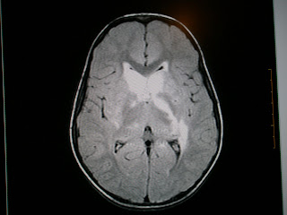
Acute disseminated encephalomyelitis (ADEM) is an immune mediated disease of brain. It usually occurs following a viral infection but may appear following vaccination, bacterial or parasitic infection, or even appear spontaneously. It involves autoimmune demyelination.Full recovery is seen in 50 to 75% of cases, ranging to 70 to 90% recovery with some minor residual disability, with an average time to recover of one to six months.MRI is highly sensitive in detecting white matter lesions and the lesions described are rather extensive and subcortical in location. Involvement of the deep gray matter, particularly basal ganglia, is more frequent. Use of high-dose methylprednisolone, plasma exchange, and IVIG are based on the analogy of the pathogenesis of ADEM with that of multiple sclerosis (MS). Differentiation of ADEM from the first attack of MS is important from prognostic as well as therapeutic point of view. This differentiation is more relevant to India where the incidence of MS is low.
To read more: click here
Dermoids Here and There
Intracranial Dermoids:
Most dermoid tumors have signal intensity characteristics similar to fat—that is, they are hyperintense on T1-weighted images and hypointense on T2-weighted images. Fat-suppression techniques may be helpful in confirming the presence of fat in the lesion. Centrally, dermoid tumors may appear inhomogeneous due to the presence of hair follicles, calcifications, and cellular debris.Rupture of a dermoid tumor can result in fat droplets in the subarachnoid spaces or ventricles.
Spinal Dermoids:
The most frequent site of these tumors is in an extramedullary location; a few may lie in the substance of the cord itself. Complications of the cyst include secondary infection, particularly when associated witha dermal sinus. Rarely these lead to chemical meningitis from the cholesterol-laden keratin released in the subarachnoid space from a ruptured cyst leading to a foreign body type of reaction.
Presacral Dermoid:
A unilocular cystic mass located at or near the midline, with signal intensity similar to that of fat, is highly suggestive of a dermoid tumor.
Submitted by Dr MGK Murthy (Elbit Medical Diagnostics)
Chordoma
Large expansile infiltrative lesion in the S1 and S2 vertebral bodies with pre and paravertebral and epidural components with consequent neural compromise and spinal canal narrowing.
Chordomas are rare tumors that arise from embryonic notochordal remnants along the length of the neuraxis at developmentally active sites.Chordomas are rare neoplasms. As primary intracranial neoplasms, they only constitute 0.2% of all CNS tumors. Chordomas generally occur in 3 locations, which are, in descending order of frequency, the sacrum, intracranially at the clivus, and along the spinal axis.CT scan or MRI studies are indicated to evaluate the extent of the tumor and to identify the tissues that the chordoma has infiltrated.The 5-year survival rate is estimated to be 51%, and the 10-year survival is estimated to be 35%. Factors that may improve prognosis are young age, complete resection, and the addition of radiation therapy in incompletely resected tumors.
You can find more information on this topic here
Saturday, March 8, 2008
Coexistent Carcinomas
Known case of prostatic carcinoma with increased PSA levels and osteoblastic metastasis in the spine. Interestingly this patient also had para-aortic adenopathy and a testicular swelling. This could well represent metastatic lymphadenopathy but the question of coexistent prostatic and testicular carcinomas should be raised. The incidence of coexistent malignancies is high in the elderly.
Malignant chacracteristics of a breast lesion
MRI - Sensitivity - 93% Specificity - 70%
1. Opacity
2. Enhancement pattern : patchy
- invasive: pheripheral or rim
- DCIS : linear
3. Type 3 time intensity curve
- early steep rise within 5 mts with a 70% increase in SI
- rapid washout
4. Tumour recurrence – radiation fibrosis does not enhance after 18 mts
Ultrasound :
Poorly reflective
Ill defined
Heterogenous internal echo pattern
Absent farwall echoes
Posterior acoustic shadowing
Taller than wide
Mammography:UOQ - 50% Retroareolar 18%
Opacity - stellate,spiculated,comet tail
- high density
- nonhomogenous
- wide halo
Asymmetric Density
Microcalcification – 30% of cases
- < 0.5 mm & > 5 particles per cc
- segmental, cluster distribution
- pleomorphic : linear, branching, punctate
- eccentric location in opacity
Tuesday, February 19, 2008
Local Gigantism of Right Ankle and Foot
Large diffuse irregular hypertrophy of the soft tissues involving the distal leg, ankle and foot on the right side. No evidence of phleboliths or signs of arteriovenous fistulae or fat elements. The differential here for local gigantism would include Klippel Trenaunay Syndrome and Plexiform Neurofibromatosis.
Klippel-Trenaunay syndrome (KTS) is defined by the presence of a combined vascular malformation of the capillaries, veins, and lymphatics, congenital venous abnormalities, and limb hypertrophy. Most patients with KTS can be treated conservatively with compression stockings or pneumatic pumps. Compression stockings decrease edema, act as a barrier for minor trauma, and reduce venous insufficiency.
Surgical Care - Servelle reported successful surgical intervention (resection or ligation of abnormal blood vessels) in more than 700 patients with KTS. Most medical centers have tried to avoid surgical intervention. Surgical treatment can be complicated by infection, lymph seepage, and skin breakdown. Intravenous sclerotherapy has been proposed as an alternative to surgical intervention in KTS and to embolization in PWS. Reports exist of the use of a sclerosant in microfoam.
Clinicians at all centers agree that a leg length discrepancy of more than 2.0 cm warrants epiphysiodesis. Hypertrophied digits with severe deformity and infection may require amputation.
You can find more information on this topic here
Posterior Reversible Encephalopathy Syndrome (PRES)
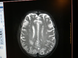
Multiple illdefined irregular altered signal intensity areas involving both parietoccipital regions in parasagital location.
Posterior Reversible Encephalopathy Syndrome (PRES) or Reversible Posterior leukoencephalopathy syndrome (RPLS) is an increasingly recognized neurologic disorder with characteristic computed tomographic (CT) and magnetic resonance (MR) imaging findings, and it is associated with a multitude of diverse clinical entities. These include acute glomerulonephritis, preeclampsia and eclampsia, systemic lupus erythematosus, and thrombotic thrombocytopenic purpura and hemolytic-uremic syndrome, as well as drug toxicity from agents such as cyclosporine, tacrolimus, cisplatin, and erythropoietin. Most, but not all, cases manifest with acute to subacute hypertension, and seizures are also frequent. Classic CT findings are those of bilaterally symmetric low attenuation in the posterior parietal and occipital lobes, whereas MR imaging demonstrates hyperintensity on T2-weighted images in the same distribution.
Two pathophysiologic mechanisms for RPLS have been proposed. One postulates cerebral vasospasm with resulting ischemia within the involved territories, whereas the other posits a breakdown in cerebrovascular autoregulation with ensuing interstitial extravasation of fluid. Diffusion MR imaging can be used to discriminate between these two possibilities, as the cytotoxic edema of cerebral ischemia demonstrates decreased water mobility, whereas vasogenic edema due to cerebrovascular autoregulatory dysfunction results in increased water mobility.
You can find more information on this topic here
Lateral Medullary Syndrome
Acute infarct on the lateral aspect of medulla with extension into the superior cerebellar peduncle.
Lateral medullary syndrome (also called Wallenberg's syndrome and posterior inferior cerebellar artery syndrome) is a disease in which the patient has difficulty with swallowing or speaking or both owing to one or more patches of dead tissue (known as an infarct) caused by interrupted blood supply. It is the clinical manifestation resulting from occlusion of the posterior inferior cerebellar artery (PICA) or one of its branches or of the vertebral artery, in which the lateral part of the medulla oblongata infarcts, resulting in a typical pattern.
In contrast Medial medullary syndrome (also known as Dejerine syndrome) is a set of clinical features resulting from an infarction in the brainstem. The vessel usually involved is the anterior spinal artery which supplies the medial part of the medulla oblongata.
You can find more information on this topic here
Right Acoustic Neuroma
Right extraxial cerebellopontine angle lesion with a tongue of tissue extending into the internal audirary canal.
An acoustic neuroma, also called a vestibular schwannoma, is a benign primary intracranial tumor of the myelin-forming cells of the vestibulocochlear nerve (CN VIII).Acoustic neuromas may occur sporadically, or in some cases occur as part of von Recklinhausen neurofibromatosis, in which case the neuroma may take on one of two forms. In Neurofibromatosis type I, a schwannoma may sporadically involve the 8th nerve, usually in adult life, but may involve any other cranial nerve or the spinal root. In Neurofibromatosis type II, bilateral acoustic neuromas are the hallmark and typically present before the age of 21. These tumors tend to involve the entire extend of the nerve and show a strong autosomal dominant inheritance. Contrast-enhanced CT will detect almost all acoustic neuromas that are greater than 2.0 cm in diameter and project further than 1.5 cm into the cerebellopontine angle. Those tumors that are smaller may be detected by MRI with gadolinium enhancement. Audiology and vestibular tests should be concurrently evaluated using air conduction and bone conduction threshold testing to assess for sensorineural versus conduction hearing loss. Indicated treatments for acoustic neuroma include surgical removal and radiotherapy.
You can find more information on this topic here
Bilateral femoral AVN
Both femoral heads show the crescent sign with altered signals in the left femoral neck and free fluid in the joints (left > right)
Pathologic changes leading to AVN are initiated in two broad categories of anatomic regions, intravascular(arterial and venous)and extravascular factors (intraosseous and capsular). Diseases or conditions associated with or leading to AVN are Truama, Hemoglobinopathies, Polycythemia, Caisson disease - Dysbaric osteonecrosis and Gaucher's disease, Infection, Neoplasms, Hypercortisolism - Corticosteroid medications and Cushing's disease, Alcohol consumption, Pancreatitis, Chronic renal failure, Cigarette smoking, Collagen vascular diseases, Congenital and developmental, Organ transplantation.
MRI is the most sensitive means of diagnosing AVN. MRI provides the criterion standard of noninvasive diagnostic evaluation. It is more sensitive than CT scanning or planar scintigraphy, and is much more sensitive than plain film radiography for detecting AVN.A staging system using radiographic findings has been developed by Ficat and Arlet and has been used widely for treating AVN. This has been supplanted by the classification system of Steinberg et al, which incorporates MRI and scintigraphic findings.
You can find more information on this topic here
Friday, January 25, 2008
Glomus Jugulare
Large expansile grossly destructive irregular space occupying lesion involving the left temporal bone with intracranial extension and mass effect on brain stem, fourth ventricle and temporal horn.
Glomus jugulare tumors are rare, slow-growing, hypervascular tumors that arise within the jugular foramen of the temporal bone. They are included in a group of tumors referred to as paragangliomas, which occur at various sites and include carotid body, glomus vagale, and glomus tympanicum tumors.
The female-to-male ratio is 3-6:1. Glomus jugulare tumors have been noted to be more common on the left side, especially in females. The Glasscock-Jackson and Fisch classifications of glomus tumors are widely used. The Fisch classification of glomus tumors is based on extension of the tumor to surrounding anatomic structures and is closely related to mortality and morbidity.
Surgery is the treatment of choice for glomus jugulare tumors. Surgical approach depends on the localization and extension of the tumor. Intraoperative monitoring including EEGs and somatosensory-evoked potentials (SSEPs) are routinely used.Gross total resection of some extensive tumors may be extremely difficult and may carry unwarranted risk. In such cases, radiotherapy may be indicated to treat residual tumor following subtotal resection.
Submitted by Dr VN Goud (Elbit Medicla Diagnostics)
You can find more information on this topic here
Subscribe to:
Posts (Atom)